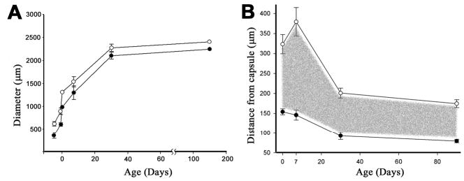
Fig. 10.

Developmental dynamics of the syncytial region in the mouse lens. (A) The equatorial diameter of the lens (open symbols) and the uniformly labeled syncytial region (closed symbols) were measured in embryonic and adult lenses. The region expanded during development, closely matching the overall growth rate of the lens. (B) The thickness of the variegated region (filled symbols) decreased during early postnatal development before stabilizing at approximately 90 μm in adult lenses. The thickness of the nucleated cell layer (open symbols) also stabilized in older animals at approximately 180 μm. The layer of nucleated cells within the syncytial region (indicated by the shaded area) was approximately 90 μm thick. Data points represent the mean±s.d. (n>8 lenses for each time point).
