
Fig. 4.
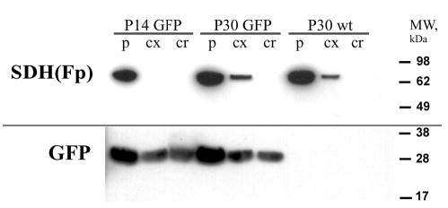
GFP proteolysis does not occur during fiber cell differentiation. Lenses from P14 or P30 TgN(GFPU)5Nagy mice or wild-type controls were separated into three regions. The peripheral sample (p) contained the region of variegated GFP expression. The core sample (cr) was from the center of the lens and contained cells of uniform fluorescence. A cortex sample (cx) was obtained from a region intermediate between the peripheral and core samples. Blotted samples were probed with antibodies against the mitochondrial marker SDH(Fp) or GFP (see text for details). SDH(Fp) was abundant in the peripheral samples from transgenic or wild-type lenses, reduced in the cortex and absent from the core. The GFP antibody identified a single band of the expected size (27 kDa) in samples from the TgN(GFPU)5Nagy lenses. This band was absent from the wild-type controls. Full-sized GFP was present in samples from each region of the transgenic lenses, including the oldest cells in the lens, those of the core. No evidence of proteolytic breakdown products was observed. The results are representative of three experiments.