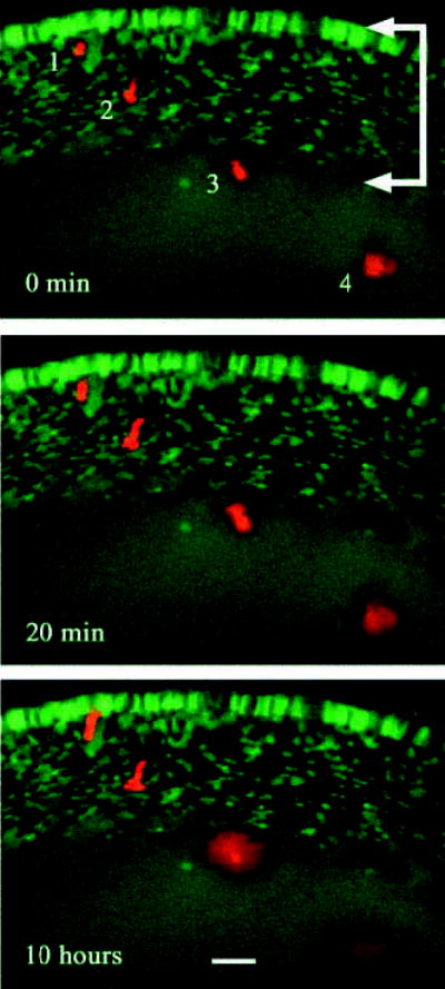
Fig. 6.

Intercellular diffusion of 10 kDa TMRD occurs only in the uniformly labeled core region of TgN(GFPU)5Nagy hemizygous mouse lenses. Four fiber cells located at various depths below the surface of a P2 transgenic lens were injected with TMRD (red). Intrinsic GFP fluorescence (green) and TMRD fluorescence were covisualized during a 10 hour period in organ culture. Cell #1 and cell #2 were located in the variegated layer (arrows) at the lens surface. In these cells, TMRD fluorescence was retained over the course of the experiment. Cell #3 and cell #4 were located deeper below the lens surface. Cell #3 lay just within the region of uniform GFP fluorescence and cell #4 was located approximately 80 μm within the uniform region. Over the 10 hour culture period, TRMD fluorescence was not retained by cell #3 or cell #4. At the end of the experiment, TMRD fluorescence was visible as a diffuse cloud that had spread well beyond the boundaries of the injected cells. Bar, 25 μm.
