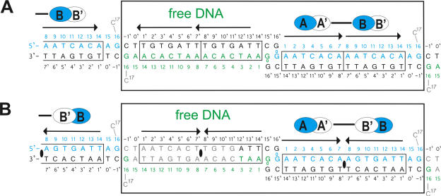
Figure 1.
DNA used for cocrystallization and arrangement of (A) [Δ19ω2]2-(→→) and (B) [Δ19ω2]2-(→←) and free DNAs in the crystal asymmetric units (large boxes). Subunits sharing the same protein–DNA interactions Δ19ωA/B shown in blue and Δ19ωA′/B′ in white ovals. Solid bars indicate subunits interacting through helices α1, α1′. The pseudo-2-fold axis relating Δ19ω2 and bound heptads in [Δ19ω2]2-(→←) indicated by black ellipses in (B). Heptads with sequence 5′-AATCACA/T-3′ outlined by arrows, and nucleotides are numbered 0 to 17 and −1′ to 16′, respectively. Top strands of free DNA in green. Grey nucleotides C17 in both structures and 4 to 15 bp of free DNA in [Δ19ω2]2-(→←) could not be modeled due to poor electron density.

