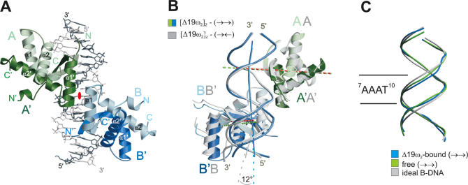
Figure 2.
(A) Structure of [Δ19ω2]2-(→→). DNA backbone trace in light grey for top strand and dark grey for bottom strand, see Figure 1. Δ19ω monomers A/A′ and B/B′ in light and dark green and blue, respectively, helices α1 and α2 labeled with white letters. Helices α1′ of A′ and α1 of B related by a pseudo-2-fold axis perpendicular to the paper plane (red ellipse) form the Δ19ω2···Δ19ω2 interface. (B) Superimposition of Δ19ωA/A′ of [Δ19ω2]2-(→→) (green) and Δ19ωA/A′ of [Δ19ω2]2-(→←) (grey) to show slight positional differences of Δ19ω B/B′ associated with palindromic symmetry in (→←). Pseudo-2-fold axes relating monomers in Δ19ω2 indicated by dashed lines colored green for [Δ19ω2]2-(→→) and red for [Δ19ω2]2-(→←). Helices α1, α1′ involved in Δ19ω2···Δ19ω2 interactions superimpose well allowing cooperative binding in both complexes. DNA in [Δ19ω2]2-(→←) is locally kinked by 12° at the centre (bp G11–C4′) of heptad A8–T14, and Δ19ω2 are ∼0.6 Å closer (vertical distance between pseudo-2-fold axes) than in [Δ19ω2]2-(→→), see text. (C) Phosphate backbone of the 14 bp operator regions of operator DNA free (→→) in green and Δ19ω2-bound (→→) in blue superimposed on ideal B-DNA (grey).

