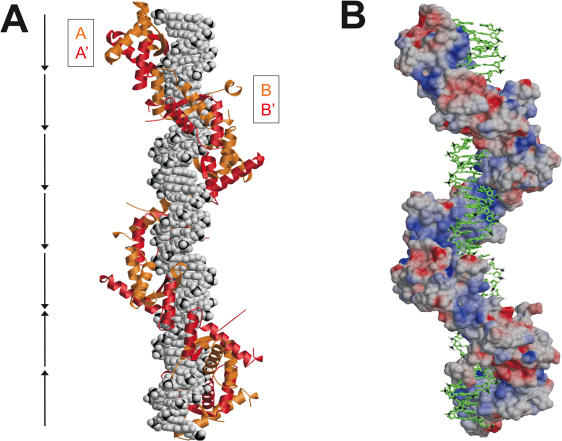
Figure 8.
Model of seven Δ19ω2-bound heptads (→→→→→←←) of the natural promoter Pδ. (A) DNA in space filling and Δ19ω2 as orange/red ribbons, (B) DNA in stick and Δ19ω2 in surface representation colored according to electrostatic potential (negative and positive charges red and blue, respectively). The model is based on the structures of [Δ19ω2]2-(→→) and [Δ19ω2]2-(→←). Repressors form a left-handed protein-matrix winding around the nearly straight operator.

