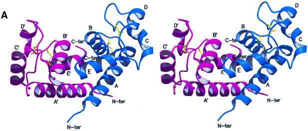
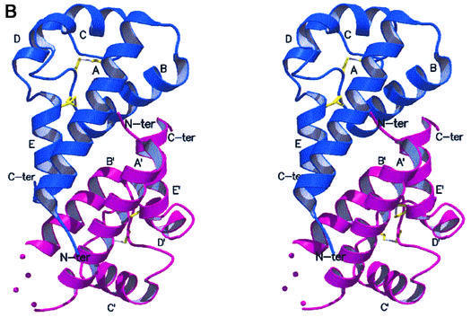
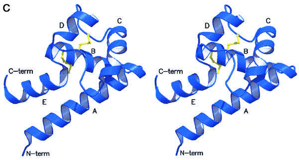
Fig. 2. Stereoview of the dimeric hCD81-LEL. The view, generated with MOLSCRIPT and RASTER3D (Kraulis, 1991; Merritt and Murphy, 1994), shows the two subunits in blue and purple. The helix labels are distinguished in the two protomers by a prime. The molecular 2-fold axis is close to vertical (A) and located between the N- and C-termini of the two chains. Solid dots (in the purple subunit) trace an approximate path for residues 238–241 not observed in the electron density maps. In (B), the 2-fold axis is orthogonal to the drawing plane. (C) Stereoview of the hCD81-LEL isolated protomer. The view highlights the head subdomain localization relative to the N- and C-terminal helices (α-helices A and E, respectively), and the labeling of secondary structure elements. The two disulfide bridges are shown in yellow.

An official website of the United States government
Here's how you know
Official websites use .gov
A
.gov website belongs to an official
government organization in the United States.
Secure .gov websites use HTTPS
A lock (
) or https:// means you've safely
connected to the .gov website. Share sensitive
information only on official, secure websites.


