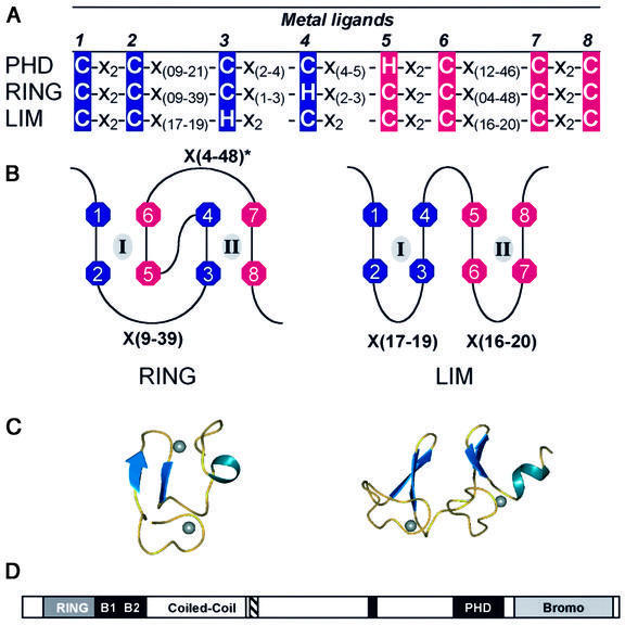
Fig. 1. (A) The consensus sequences that define the PHD, RING and LIM domains. C, cysteine; H, histidine; X, any residue. Above the consensus sequence the number of each metal ligand, ml, is given. The first and second pairs of sequential metal ligands are in dark blue, while the third and fourth pairs are in magenta. (B) Demonstration of zinc ligation patterns found in RING and LIM domains. The RING uses a cross-brace ligation scheme while the LIM uses a sequential ligation scheme. Numbers correlate to the number of the metal ligand as defined in (A). Zinc atoms are represented by gray ovals and the zinc-binding sites are denoted by roman numerals. (C) Ribbon diagrams of the three-dimensional structures. Zinc atoms are represented by spheres. Structures are from the RING of PML (1BOR) and for LIM (1A7I). (D) A schematic representation of the domains in the KAP-1 protein. RING, the RING domain; B1 and B2, two adjacent B-boxes; Coiled-Coil, a leucine coiled-coil domain; hatched box, the TIFF signature sequence; black box, the HP1-binding site; PHD, the PHD domain; Bromo, the bromodomain.

An official website of the United States government
Here's how you know
Official websites use .gov
A
.gov website belongs to an official
government organization in the United States.
Secure .gov websites use HTTPS
A lock (
) or https:// means you've safely
connected to the .gov website. Share sensitive
information only on official, secure websites.
