
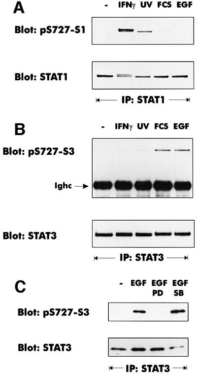
Fig. 6. Different signaling pathways cause S727 phosphorylation of STAT1 and STAT3. Where indicated, mouse 3T3 fibroblasts were treated with IFN-γ for 30 min, irradiated with UV light, or stimulated with 10% FCS (30 min) or EGF (100 ng/ml, 30 min). (A) STAT1 phosphorylation on S727 was analyzed with phosphospecific antiserum. Loading was controlled by reprobing with a monoclonal antibody to the STAT1 N-terminus. (B) STAT3 was precipitated from cellular lysates with an antiserum to the C-terminus. Phosphorylation of S727 was determined with a phosphoserine 727-specific antiserum (Ighc indicates the immunoglobulin heavy chain). The loading control was performed by reprobing the blot with the STAT3 C-terminal antiserum. (C) The pathway-specific inhibitors PD98059 (PD) and SB203580 (SB) were added 30 min prior to EGF treatment. STAT3 phosphorylation on S727 was determined as described for (B).