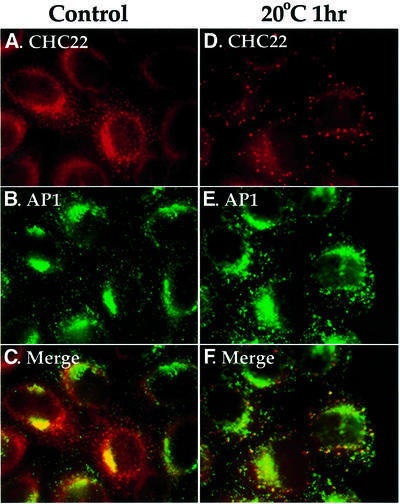
Fig. 6. Disruption of CHC22 TGN localization by low temperature. HeLa cells, infected with pLNCXCHC22 to obtain low-level enhanced expression of CHC22, were either left untreated (A–C) or incubated at 20°C for 1 h (D–F) before being subjected to digitonin wash to remove cytosol, and then processed for immunofluorescence. CHC22 (red) was detected with anti-CHC22 antiserum followed by LRSC-conjugated goat anti-rabbit IgG (A and D). AP1 (green) was stained with MAb 100/3 followed by FITC-conjugated goat anti-mouse IgG (B and E). The columns represent the same images viewed with different filters and the bottom panels are merged images.

An official website of the United States government
Here's how you know
Official websites use .gov
A
.gov website belongs to an official
government organization in the United States.
Secure .gov websites use HTTPS
A lock (
) or https:// means you've safely
connected to the .gov website. Share sensitive
information only on official, secure websites.
