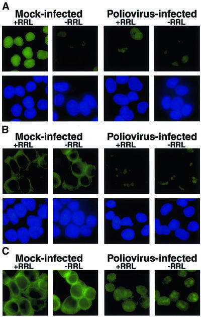
Fig. 4. Cell-free nuclear import assays. (A) Uninfected cells (Mock-infected) or cells that had been infected with poliovirus for 4 h (Poliovirus-infected) were permeabilized and used in an in vitro nuclear import assay. Assays were carried out in the presence (+RRL) or absence (–RRL) of rabbit reticulocyte lysate as a source of cytosolic factors. Top panels show GFP using a FITC filter and bottom panels show Hoechst 33258 staining of DNA using a UV filter. All GFP images were acquired using identical exposure times and manipulations in Photoshop 5.0. (B) Same as in (A), except that creatine kinase, creatine phosphate, ATP and GTP were omitted from the reaction. GFP images were acquired using the same exposure time as in (A). (C) Longer exposure of GFP fields in (B).

An official website of the United States government
Here's how you know
Official websites use .gov
A
.gov website belongs to an official
government organization in the United States.
Secure .gov websites use HTTPS
A lock (
) or https:// means you've safely
connected to the .gov website. Share sensitive
information only on official, secure websites.
