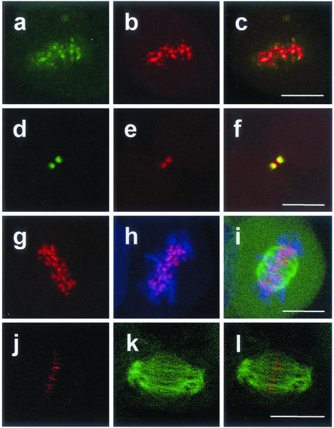
Figure 3.
SurvivinDsRed colocalizes with Aurora B-kinase and with the ends of microtubules at kinetochores and the central spindle. Analysis by confocal laser scanning microscopy. a–f, HeLa cells transduced with 5 MOI pcz-CFG5.1-survivinDsRed and stained with an anti-AIM-1/Aurora B-kinase antibody in indirect FITC-immunofluorescence. a, b, and c, metaphase. a, Aurora-B kinase, b, survivinDsRed; c, merge of a and b. Note that Aurora-B kinase and survivinDsRed are colocalized at kintetochores. d–f, late telophase. d, Aurora-B kinase, e, survivinDsRed; f, merge of d and e. Note that AIM-1/Aurora-B-kinase and survivinDsRed are colocalized at the midbody. g–l, HeLa cells were double transduced with 5 MOI of pcz-CFG5.1-survivinDsRed and 10 MOI of pcz-CFG2-EGFP-tubulin and stained with TO-PRO-3 dye. g, survivinDsRed. h, survivinDsRed/TO-PRO-3 merge. i, survivinDsRed/TO-PRO-3/EGFP-α-tubulin triple merge. j–l, late anaphase/early telophase. HeLa cells were double transduced with 5 MOI of pcz-CFG5.1-survivinDsRed and 10 MOI pcz-CFG2-EGFP-α-tubulin. j, survivinDsRed; k, EGFP-α-tubulin; l, merge of j and k. Note that survivinDsRed is colocalized with the ends of the polar microtubules. Bar (a–i), 10 μm; (g–l), 15 μm.

