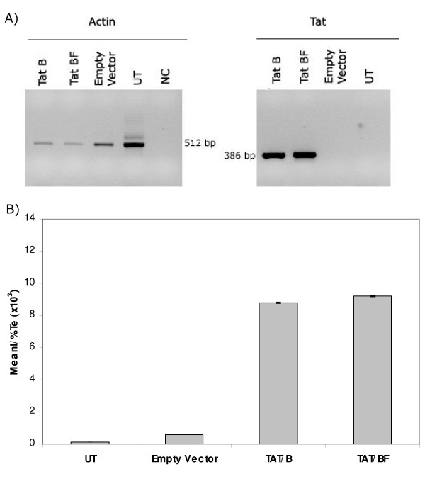
Figure 5.
Transactivation of GFP expression in GHOST cells. A) tat mRNA was detected in pTat transfected cells, but not in untransfected cells or cells transfected with empty vector, by RT-PCR (right panel). Amplification of actin mRNA was also evaluated as an internal control (left panel). B) Tat expressing vectors were capable to transactivate the expression of the gfp gene placed downstream the LTR from HIV-2. No statistical difference was found between TatB(NL4-3) and TATBF(ARMA159). UT: Untransfected cells, NC: Negative control, MeanI: Mean Fluorescence Intensity, Te: Transfection efficiency. Data presented here is representative of 4 independent assays.

