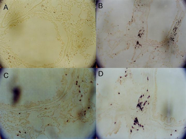
Figure 3.

Immunohistochemical examination for CD8+ T cells in lung tissue. In the lung tissue from the vaccination group, more CD8+ T cells were infiltrated along the airway than in control group. A and B, Lung tissue from control group mouse (×100 and ×200). C and D, Vaccination group (×100 and ×200). Immunohistochemical staining with rat anti-mouse CD8 monoclonal antibody.
