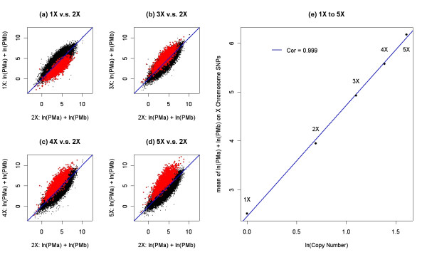
Figure 1.
Panels a-d show the standardized ln(PMa) + ln(PMb) intensity for the 1X, 3X, 4X, and 5X DNA samples relative to the intensity of the 2X DNA sample. Black data points correspond to autosomal SNPs and red data points correspond to the 1,955 X-chromosome SNPs. The blue line in each panel represents the Y = X line. Panel e shows the relationship between the natural log-transformed copy number and the natural log-transformed intensity. The x-axis is the natural log-transformed copy number and the y axis is the average ln(PMa) + ln(PMb) intensity across 1,955 SNPs. The blue line is the regression using the average intensity as the response and the natural log-transformed copy number as the predictor.

