
Fig. 1.
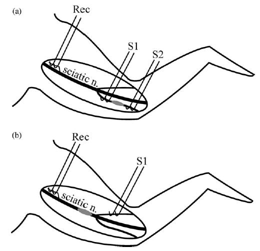
Diagram of electrophysiology setup in animals with (a) a peroneal nerve and (b) a sciatic nerve lesion. In (a) peroneal nerve fibres are stimulated proximal (S1) and distal (S2) to the lesion site (grey ellipse). In (b) sural nerve fibres are stimulated distal (S1) to the lesion site (grey ellipse). In both preparations, recordings are made from fine filaments teased from the sciatic nerve proximal to the lesion site. S1: stimulus 1, S2: stimulus 2; Rec: recording electrodes.