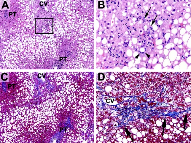
FIGURE 1. Representative histologic sections of a liver biopsy before bariatric surgery. A, Most preoperative livers contained abundant steatosis. In this example (H&E stain), macrovesicular steatosis is present throughout most of the lobules with relative sparing of the periportal hepatocytes (PT, portal tract; CV, central vein). B, Many preoperative livers also had evidence of steatohepatitis. The boxed area seen in (A) is shown here. Several hepatocytes are surrounded by inflammatory cells (one such hepatocyte is indicated by the arrowheads). The inflammatory infiltrate is composed of neutrophils (arrows), lymphocytes and macrophages (arrowheads). C, Many preoperative livers also had increased fibrosis, as highlighted here with a trichrome stain (same section as shown in A). Pericellular fibrosis is noted around the central vein (blue strands) without significant periportal fibrosis, indicating stage 1 fibrosis (usually, the central veins are free of any fibrosis). Hepatocytes are dark red with this stain. D, Higher power view of another area showing centrilobular pericellular fibrosis (emphasized by the arrows).
