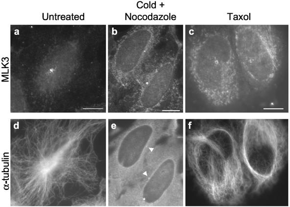
Figure 4.
Localization of the centrosome-associated MLK3 sprinkles is partially dependent on microtubules. Asynchronous HeLa cells growing on coverslips were either: 1) untreated (a and d); 2) incubated in the presence of nocodazole (6 μg/ml) first on ice for 45 min and then at 37°C for 1 h (b and e); or 3) treated with taxol (5 μM) for 4 h (c and f). Cells were fixed in methanol and stained for MLK3 (a–c) and α-tubulin (d–f). Arrowheads in e denote brighter spots of α-tubulin staining indicative of centrosomes. The reduced levels of MLK3 staining after treatment with cold/nocodazole or taxol required longer photographic exposures for imaging leading to a commiserate increase in the level of cytoplasmic staining seen in b and c relative to a. Scale bars, 10 μM, applies to all images of a given field.

