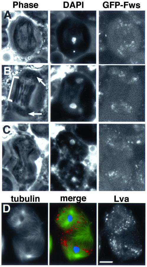
Figure 7.
Fws localization in wild-type spermatocytes undergoing meiosis I. Live spermatocytes expressing GFP-Fws and stained with Hoechst dye to visualize DNA in (A) metaphase, (B) early telophase, and (C) mid telophase. (B) Astral membranes are marked by arrows, and central spindle is marked by bracket. (D) Fixed spermatocyte in telophase stained with Lva antibodies to visualize Golgi membrane (red), anti-α-tubulin antibodies to visualize microtubules (green), and DAPI to visualize the DNA (blue). Black and white panels show anti-tubulin and anti-Lva staining, respectively. Bar, 10 μm.

