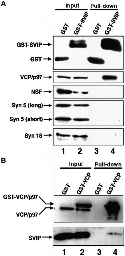
Figure 2.
Binding between VCP/p97 and SVIP. (A) GST or GST-SVIP was expressed in 293T cells and then the cells were lysed. After centrifugation, GST (lane 1) or GST-SVIP (lane 2) in the supernatant was pulled down with glutathione beads. The proteins coprecipitated with GST (lane 3) or GST-SVIP (lane 4) were detected by immunoblotting with antibodies against proteins as indicated on the left. The amount of each protein in 4.4% of the supernatant is shown (input). (B) Expressed GST (lane 1) or GST-VCP/p97 (lane 2) was pulled down with glutathione beads, and the proteins coprecipitated with GST (lane 3) and GST-VCP/p97 (lane 4) were detected by immunoblotting with antibodies against VCP/p97 and SVIP. The amounts of GST-VCP/p97, endogenous VCP/p97, and endogenous SVIP in 4.4% of the supernatant used are shown (input).

