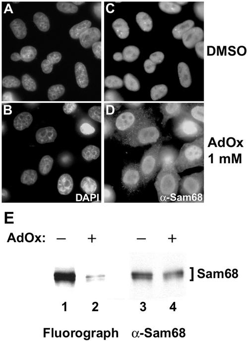
Figure 5.
Sam68 accumulates in the cytoplasm in the presence of methylase inhibitors. HeLa cells were treated with either DMSO (A and C) or with the methyltransferase inhibitor AdOx at 1 mM (B and D) for 24 h. The cells were fixed and immunostained with the anti-Sam68 AD1 antibody followed by a secondary antibody conjugated to Alexa 488 (green; C and D). Nuclei were visualized by the DNA-specific stain 4,6-diamidino-2-phenylindole (blue; A and B). (E) HeLa cells were incubated with AdOx for 24 h preceding an in vivo methylation labeling. Immunoprecipitations were performed with anti-Sam68 AD1 antibodies. Samples were separated by 10% SDS-PAGE, transferred to a polyvinylidene difluoride membrane, sprayed with Enhance, and exposed to film overnight. The 3H-labeled proteins were visualized by fluorography (left), and the same membrane was immunoblotted with anti-Sam68 AD1 antibodies (right).

