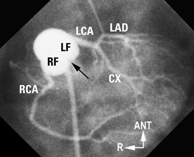
Fig. 6 “Usual” coronary artery anatomy in d-TGA. Laid-back aortogram shows a rightward anterior aorta. Both facing sinuses are well seen, as is the facing commissure (arrow). Both coronaries arise from the mid-points of their respective facing sinuses (RF, LF) and show a good length of proximal vessel before the 1st branch.
ANT = anterior; CX = circumflex; d-TGA = complete transposition of the great arteries; LAD = left anterior descending; LCA = left coronary artery; LF = left facing; R = right; RCA = right coronary artery; RF = right facing
(Modified from: Freedom RM, et al. 2 With permission of Futura Publishing Company, Inc.)
