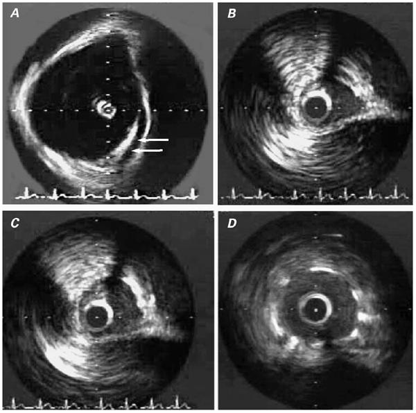
Fig. 2 Patient 2. Intravascular ultrasonographic (IVUS) images: A) Image obtained within the aortic root, illustrating the tangential, intramural course of the proximal right coronary artery (arrows). B) Intramural segment of the right coronary artery (RCA) during systole. C) Same segment as in B, during late diastole (maximal lumen). D) Intramural RCA after stent implantation.
