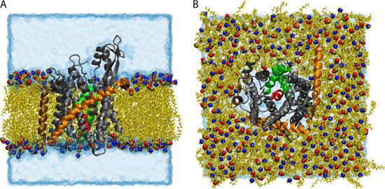
FIGURE 1.
Simulated system of SecYEβ in a lipid bilayer/water environment. SecYEβ is shown in cartoon representation with SecY, SecE, and Secβ colored in gray, orange, and ochre, respectively. The plug, transmembrane domain 2a of SecY (residues Ile55 to Gly65) is presented in red, and TM2b (residues Gly76 to Ser91) and TM7 (residues Asn256 to Gly280) are shown in green (both also of SecY). The lipids are seen in yellow licorice representation with the phosphorus, nitrogen, and an oxygen of the headgroup highlighted as spheres colored in tan, blue, and red, respectively. The water box is drawn in transparent blue surface representation. (A) Side view of the simulated system. To display the protein more clearly, some lipids and water molecules have been removed, leaving a flat outward face. (B) Top view of the simulated system. The top solvation layer has been removed.

