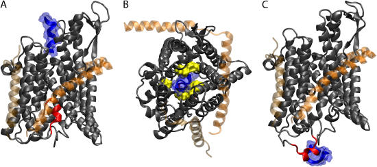
FIGURE 3.
Translocation of deca-alanine through SecYEβ. The figure shows the results of simulation sim1 (see Table 1). The representation and coloring of SecY, SecE, and Secβ is the same as in Fig. 1 with the exception that SecE and Secβ are rendered transparent. The plug (TM2a of SecY, residues Ile55 to Gly65) is shown in red. (A) Front view of SecYEβ and deca-alanine at t = 0. Deca-alanine is shown in blue cartoon representation together with its (transparent) surface and is positioned on the cytoplasmic side of SecYEβ before translocation. (B) Top view of the SecYEβ-deca-alanine system at t = 0.8 ns. The pore ring (residues Ile75, Val79, Ile170, Ile174, Ile260, and Leu406 of SecY), expanded from its equilibrium (t = 0) state, is shown in surface representation colored yellow with deca-alanine passing through it. Deca-alanine is partially unfolded at this point. (C) Final state of translocation (t = 1.4 ns). The plug has been pushed out into the solvent, and deca-alanine is seen unfolded next to the plug.

