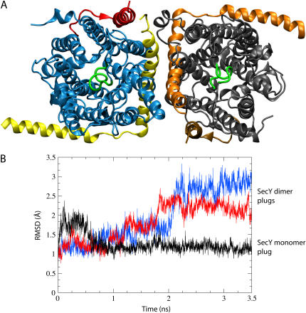
FIGURE 8.
Back-to-back dimerization of SecYEβ. (A) Dimer of SecYEβ viewed from the periplasm. Each monomer is shown in the same representation as in Fig. 1 with the exception of the color scheme of the left monomer (blue for SecY, yellow for SecE, and red for Secβ). The plugs in the dimer are without well-defined helical structure and are shown in green. (B) Destabilization of the plugs. The RMSD of the plugs (residues Ile55 to Gly65) is shown after fitting the entire SecY to the crystal structure at each point. Presented in black is the RMSD for the plug of the monomer during sim0 (see Table 1); shown in red and blue are the RMSD values for each plug from the dimer as evaluated during simulation sim5.

