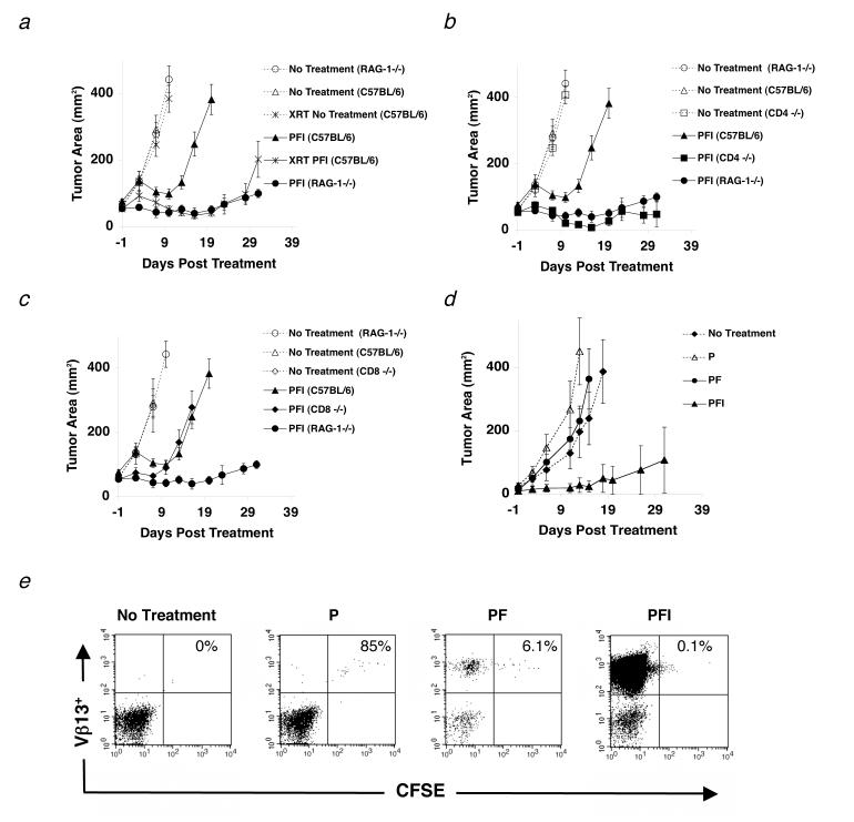
Figure 1.
Naturally occurring CD4+ T cells inhibit effective immunotherapy to established tumors. (a-d) Mice were inoculated with 1.0 × 10⁵ cells of B16 melanoma on day -14 before adoptive cell transfer with 1.0 × 10⁶ pmel-1 T cells (P), 2.0 × 10⁷ PFU rFPVhgp100 (F), and 600,000 IU of exogenous IL-2 (I), which was given daily for 3-4 days. (a) Tumor regression in C57BL/6 mice (□) is compared with RAG-1-/- mice (•) and C57BL/6 mice receiving 500 (cGy) whole body irradiation on day -1 of treatment (□). (b) Tumor regression in C57BL/6 mice (□) is compared with RAG-1-/- mice (•) and CD4-/- mice (□). (c) Tumor regression in C57BL/6 mice (□) is compared with RAG-1-/- mice (•) and CD8-/- mice (◆) in the same experiment. Data are represented as mean tumor size ± S.E.M. Experiments were independently repeated twice. (d-e) Tumor regression is IL-2 dependent. (d) Transfer of pmel-1 T cells alone (Δ) or pmel-1 T cells and rFPVhgp100 vaccine (•) into tumor-bearing RAG-1-/- hosts is similar to no treatment (◆). Addition of exogenous IL-2 with cells and vaccine is required for tumor regression (□). (e) CFSE profile of adoptively transferred pmel-1 T cells into RAG-1-/- hosts. Pmel-1 CD8+ T cells were labeled with CFSE and adoptively transferred into tumor-bearing RAG-1-/- hosts alone (P), with vaccination (PF), or with vaccination and exogenous IL-2 (PFI). 4 days later, splenocytes from treated mice were analyzed by flow cytometry. Gated on CD8+ T cells and displayed as Vβ13-PE vs. CFSE.

