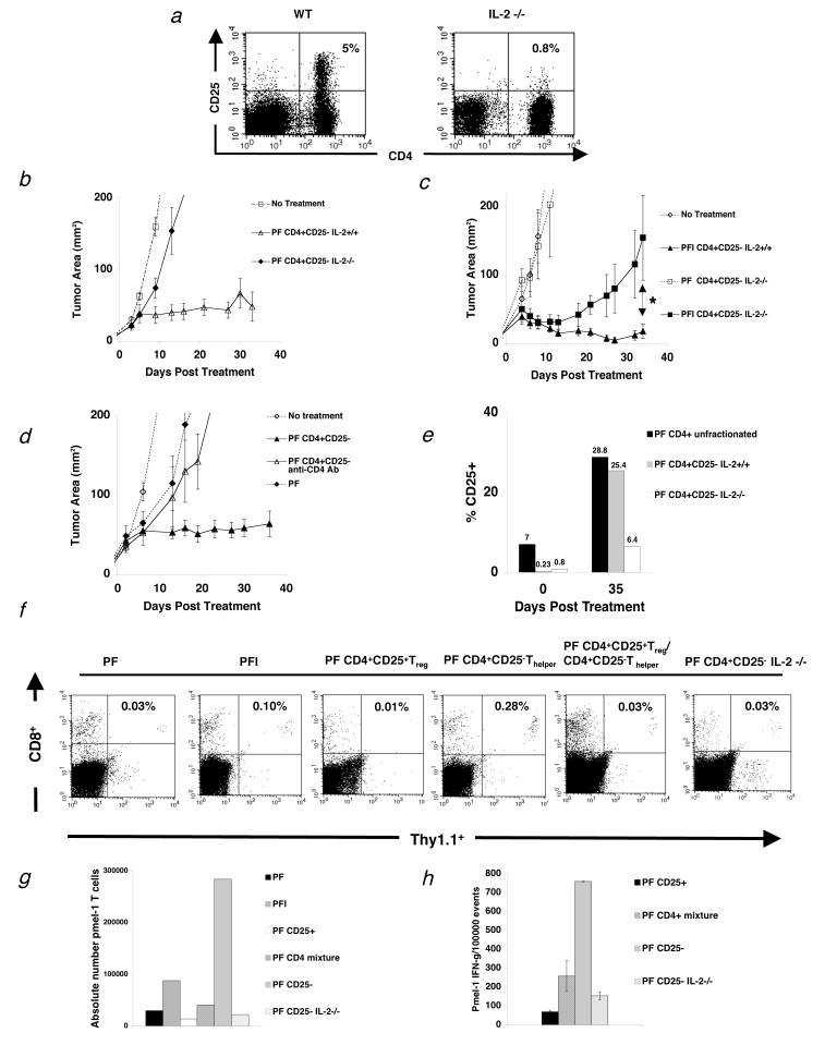
Figure 5.
T cell help is IL-2 dependent and lost in the presence of Treg cells. (a) Flow cytometry analysis of mouse splenocytes shows that IL-2-/- mice do not develop Treg cells (n=3). (b-c) RAG-1-/- mice were inoculated with 3.0 × 10⁵ cells of B16 melanoma between day -7 before adoptive cell transfer with 1.0 × 10⁶ pmel-1 T cells (P), 2.0 × 10⁷PFU rFPVhgp100 (F), plus Thelper cells from IL-2-/- mice or naturally occurring Thelper cells plus/minus exogenous IL-2 (I). (b) Transfer of Thelper cells from IL-2-/- mice with pmel-1 T cells and vaccination into tumor-bearing RAG-1-/- hosts does not help treatment of established B16 melanoma (◆). (c) Addition of exogenous IL-2 does not restore the helper function of Thelper cells from IL-2-/- mice (■). *, P = 0.021. Data are derived from a single experiment that was independently repeated 3 times. (d) Thelper cells do not program tumor-reactive CD8+ T cells. Depletion of Thelper cells 4 days after transfer with 500 mg of GK1.5 CD4 depleting mAb (Δ). Data represents 3 independent experiments with similar results. Isotype control antibody had no effect on CD4+ T cells and depletion of CD4+ T cells was confirmed by flow cytometry. (e) Thelper cells utilize IL-2 in vivo. CD25 expression on adoptively transferred Thelper cells alone, Thelper cells with Treg(CD4+unfractionated), and Thelper cells derived from IL-2-/- mice, 35 d after treatment. (f) Spleens were taken from tumor-bearing RAG-1-/- mice and analyzed by flow cytometry for the congenic marker Thy1.1 and CD8, which represents the transferred pmel-1 T cells 3 weeks after treatment with the indicated regimen. Two mice were used per group. Data is indicative of 3 independent experiments. (g) Absolute number of pmel-1 T cells 3 weeks after transfer from the same experiment in (f). (h) Intracellular IFN-γ 3 wks after adoptive cell therapy. Cells were activated with lymphocyte activating kit and analyzed by flow cytometry 6 hours later. Two mice were analyzed per group. Data is indicative of 3 independent experiments.

