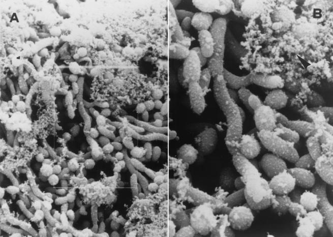
FIG. 2.
(A) Scanning electron microphotograph of blood isolate. The microphotograph shows microcolonial aggregate intimately associated with an amorphous material which seems to envelope single cells and join them (original magnification, ×950). (B) Enlargement of the image outlined in panel A (original magnification, ×2,150). The arrowhead indicates the amorphous material (slime).

