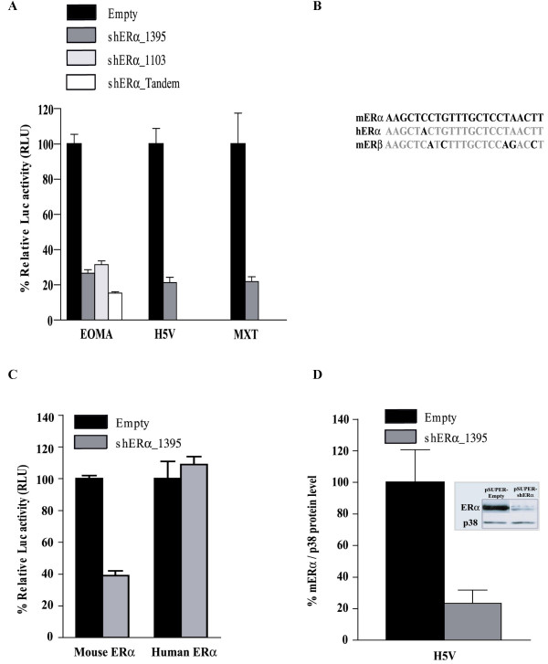
Figure 1.

Affectivity and specificity of pSUPER mediated expression of shERα in mouse cell lines. (A+C) The indicated mouse cell lines were co-transfected with, ERE-Luc, CMV-LacZ, and pSUPER-empty, pSUPER-shERα_1395, pSUPER- shERα_1103, or pSUPER- shERα_tandem. Subsequently, the cells were treated 24 hours with 10-9 M 17-β-estradiol. Luciferase activity was measured 48 hours after transfection and after correction for LacZ expression, represented as the mean (n = 3) ± SD relative to the transfection with pSUPER-empty. (A) Endogenous mouse ERα mediated transcription in EOMA, H5V and MXT cells after introducing pSUPER +/- shERα. (B) The 19-nt target-recognition sequence of ERα_1395 contains one mismatch with human ERα and five mismatches with the mouse ERβ sequence. (C) ERα mediated transcription in EOMA cells after over expression of either mouse ERα- or human ERα-expression vectors in presence of pSUPER empty or pSUPER shERα_1395 (D) Western blot analysis of H5V cells co-transfected with pCMV-mERα and pSUPER-empty or pSUPER-shERα_1395. The lysates were analysed by immunoblotting (insert-photo) with anti-mouse ERα and anti-p38. The intensity of the bands was quantified and normalized to cells transfected with pSUPER-empty. The relative ERα protein levels are presented (bar-diagram) as mean (n = 3) +/- SD.
