
Figure 1.
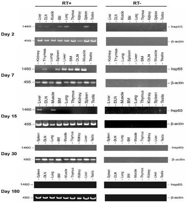
Tissue distribution of Hsp65 message. The presence of Hsp65 message in various tissue samples obtained from BALB/c mice after intramuscular immunization with 100 μg of pcDNA3-HSP65 in 25% sucrose. Total RNA (10 μg) isolated from each tissue was treated with DNase I and subjected to RT-PCR amplification with HSP65 or β-actin primers (RT+). As an RNA quality control, β-actin was amplified. No products (HSP65/β-actin) were seen when total RNA in the absence of reverse transcription was subjected to PCR amplification (RT-). All RT-PCR products were analyzed by agarose gel electrophoresis and visualized by ethidium bromide staining. The results were obtained from one mouse and are representative of three independent experiments. RT-PCR from the material obtained of mice (three animals for each point) immunized with the vector (pcDNA3) was negative in all analysis.