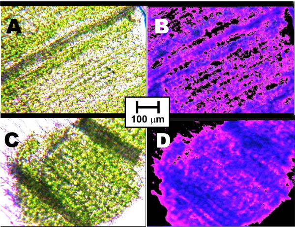
Figure 7.
Microphotos of leaf tissues. Plants were grown initially 5d etiolated, then 7d in the light. Microphotos A and B belong to sections of plants additionally exposed to Ca2+ ICR condition (BDC = 65 μT, f(BAC) = 50 Hz) during the 5d etiolation phase, while C and D are from comparable preparations of controls grown under the same conditions, but without Ca2+ ICR. A and C are taken under tungsten light, B and D under fluorescent illumination (380 nm). The plants grown at the Ca2+ ICR seemingly show a lower chloroplast density and an increased (red) fluorescence caused by free chlorophylls.

