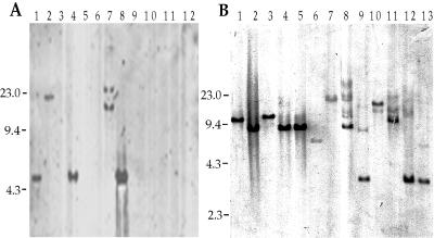
FIG. 4.
Southern analysis of the tra1 (A) and enhC (B) loci in environmental and clinical isolates of Legionella. Total bacterial DNA was digested with EcoRI and hybridized with the probes P5 against tra1 (A) and P8 against enhC (B). Minor bands in some lanes are thought to be due to partial digestion but could be due to sequences distantly related to the probe present in these strains. The strains shown are L. pneumophila AA100 (A, lane 1), L. pneumophila Allentown (A, lane 2, and B, lane 1), L. pneumophila Camperdown (A, lane 3, and B, lane 2), L. pneumophila Benidorm (A, lane 4, and B, lane 3), L. pneumophila Olda (A, lane 5, and B, lane 4), L. pneumophila Chicago 8 (A, lane 6, and B, lane 5), L. anisa (A, lane 7, and B, lane 6), L. dumoffii (A, lane 8, and B, lane 12), L. feeleii (A, lane 9, and B, lane 8), L. longbeachae (A, lane 10, and B, lane 9), L. gormanii (A, lane 11, and B, lane 10), L. pneumophila Philadelphia 1 Lp01 (A, lane 12), L. brunensi (B, lane 7), L. moravica (B, lane 11), and L. sainthelensi (B, lane 13). Numbers to the left of each panel are molecular sizes in kilobase pairs.

