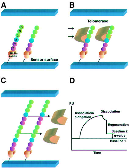
Figure 1.
Strategy for the TRE assay. (A) The TS-5′B oligomers containing the telomeric repeat sequence (TTAGGG) were immobilized on the streptavidin-pretreated dextran sensor chip surface. This gave the initial baseline on the sensorgram [baseline 1 in (D)]. (B) Telomerase extracts were injected through the flow cell and bound to the TS-5′B oligomers. Elongation of the immobilized oligomers occurred immediately. This gave the association/elongation phase on the sensorgram. (C) After finishing the injection of the telomerase extracts, dissociation of binding proteins was observed. Regeneration was performed to remove all bound proteins. (D) A sensorgram of the TRE assay. Real-time monitoring was carried out throughout the association/elongation, dissociation and regeneration phases. The difference between baselines 1 and 2 was defined as the E-value, which was equivalent to the weight of the elongated nucleotides.

