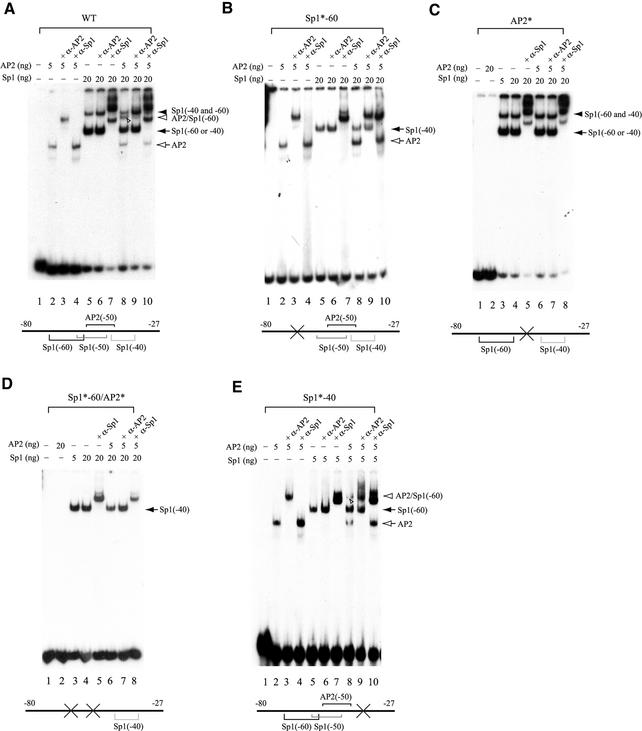
Figure 4.
Sp1 and AP2 are able to bind simultaneously to their juxtaposed binding sites in the hTAFII55 promoter-proximal region. EMSAs were performed with different amounts of purified FLAG-tagged Sp1 and FLAG-tagged AP2 proteins, in the absence or presence of anti-Sp1 (α-Sp1) or anti-AP2 (α-AP2) antibodies using labeled wild-type (WT) (A) or mutated (B–E) promoter fragments spanning –80 to –27, as indicated on each panel and described in Materials and Methods. The protein–DNA complexes detected are indicated on the right with different arrows and arrowheads. Individual protein-binding sites are marked by brackets with ‘X’ and asterisks (*) denoting mutations introduced at the specific motifs.

