
Figure 2.

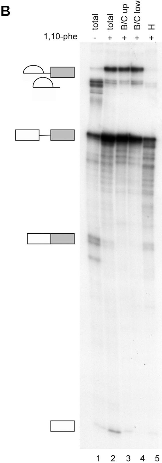

Analysis of spliceosomal complexes following the addition of 1,10-phe to the splicing reaction. (A) A radiolabeled Adeno transcript was incubated with nuclear extract under splicing conditions for the indicated duration at 30°C in the absence (–) or presence (+) of 6 mM 1,10-phe. Splicing complexes were fractionated in a 4% non-denaturing gel. (B) Analysis of the labeled mRNA contents in the B/C and H complexes. RNA from a splicing reaction incubated in the presence of 6 mM 1,10-phe for 60 min [(A), +1,10-phe; 60 min; B/C] was eluted from the gel, and the labeled RNA was extracted and separated in 8% denaturing gel. Complex B/C was divided into upper (B/C up) and lower (B/C low) parts according to the location in the gel. The RNA from complex B/C of the upper and lower parts was analyzed together with total RNA from a splicing reaction incubated in the absence (–) or presence (+) of 6 mM 1,10-phe. RNA intermediates and products are schematically represented to the left of the gel. (C) The relative intensity of the splicing complex B/C formed in the absence (squares) or presence (circles) of 1,10-phe as a function of time. Minimum and maximum intensities were normalized to zero and 100, respectively.