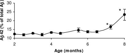
Fig. 4.
Tg brain Aβ42 levels. Quantitative analysis of Aβ42 and Aβ40 levels extracted from Tg mouse brain. Brain Aβ42 and Aβ40 was measured by using sandwich ELISA in mice from 2- to 8-month-old animals. Statistical analysis indicates that the percentage of Aβ42 rises significantly after 7 months relative to total Aβ (Aβ40 and Aβ42) levels (∗, P < 0.05).

