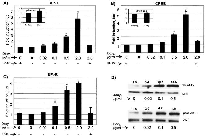
FIG. 4.
vGPCR activates AP-1, CREB, and NF-κB in PEL cells. BC-3.14 cells were transfected with the appropriate luciferase reporter and plated with the doses of doxycycline shown with or without IP-10 (100 nM), an inverse agonist of vGPCR. At 48 h, lysates were assayed for luciferase activity. (A) Dose-response curve of AP-1 activation. The inset shows the response to doxycycline of the negative control plasmid pTA-luc for the AP-1 and NF-κB constructs. (B) CREB activation. The inset shows the negative control plasmid, pFC2-dbd. (C) NF-κB activation. The results shown represent at least three independent transfections. Values are normalized to the no-doxycycline point. (D) vGPCR causes the phosphorylation of IκBα and AKT. Cells were subjected to the doses of doxycycline shown for 48 h, after which lysates (40 μg) were transferred to PVDF, probed for phosphorylated enzyme, stripped, and probed for total enzyme. Numbers above the bands represent the average fold induction (two independent experiments) in band intensity relative to baseline. ∗, In panels A to C, P ≤ 0.05 relative to the baseline uninduced value.

