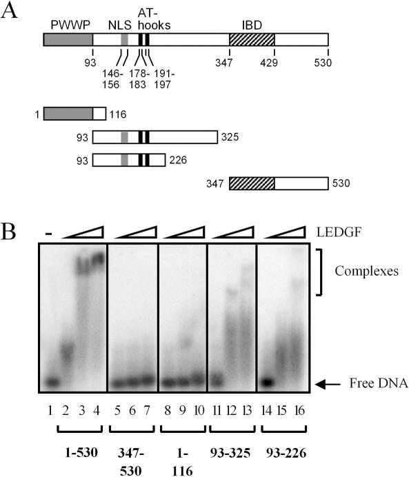
Figure 2.
LEDGF/p75 residues 93–226 harbor DNA-binding activity. (A) Schematic representation of wild-type LEDGF/p75 and deletion mutant proteins. Conserved sequence motifs/functional domains are indicated above the protein, with boundaries indicated below. NLS, nuclear localization signal; IBD, integrase-binding domain. (B) DNA-binding activities of deletion mutant proteins. DNA (100 nM Mut1) was reacted with the following protein concentrations: 250 nM (lanes 2, 5, 8, 11 and 14), 2 µM (lanes 3, 6, 9, 12 and 15) or 5 µM (lanes 4, 7, 10, 13 and 16). LEDGF was omitted from the reaction in lane 1. The results are representative of four independent gel shift experiments. The migration positions of free DNA and nucleoprotein complexes are indicated.

