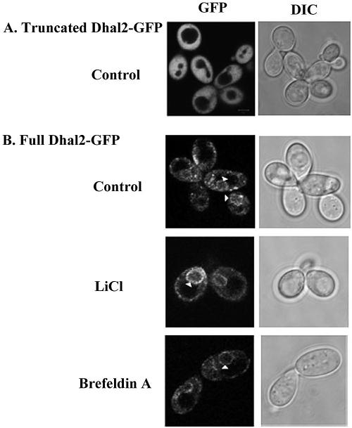
FIG. 5.
Subcellular localization of Dhal2p by fluorescence microscopy. (A) Localization of truncated Dhal2-GFP fusion protein. Strain RS16 expressing the fusion protein was grown to logarithmic phase in SD minimal medium, and GFP fluorescence was viewed under a microscope. (B) Localization of full-length Dhal2-GFP fusion protein. Strain RS16 expressing the fusion protein were grown to logarithmic phase. Cells were either untreated (control) or treated with LiCl or brefeldin A as indicated prior to visualization (see Materials and Methods). The arrow indicates the perinuclear localization of full-length Dhal2-GFP fusion protein as a ring-like structure. Bar, 5 μm.

