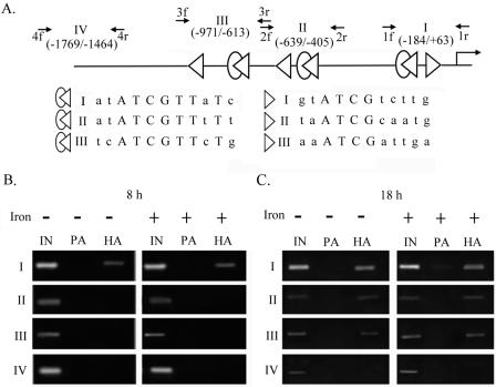
FIG. 7.
Differential promoter selection of HA-Myb1 in transfected T. vaginalis cells. The high-affinity (open merged ovals and triangles) and low-affinity (open triangles) Myb1 binding sites in the entire ap65-1 promoter are depicted in panel A, and the consensus sequences are aligned in capital letters. Small arrows indicate primer pairs used for PCRs to amplify DNAs spanning regions I, II, III, and IV of the ap65-1 promoter. The boundaries of each region relative to the transcription start site are indicated in parentheses. Samples from pFLPha-myb1/TUBneo-transfected cells under iron-depleted (left panels) or iron-replete (right panels) conditions for 8 h (B) and 18 h (C) were evaluated by a ChIP assay. PCR amplifications were performed using 1 μl of 50×-diluted DNA from input (IN) samples or 4×-diluted DNA from protein A (PA)- or anti-HA-treated (HA) samples as a template for 30 cycles. The PCR products were assayed by agarose gel electrophoresis with ethidium bromide staining.

