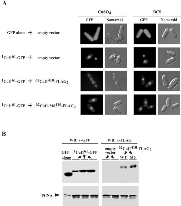
FIG. 9.
Cu-mediated inhibition of nuclear import of Cuf1 by its C-rich motif. (A) S. pombe JSY8 cells were cotransformed with an empty control plasmid and GFP alone, an empty vector and 1cuf161-GFP, 1cuf161-GFP and 62cuf1410-FLAG2, or 1cuf161-GFP and 62cuf1-M6410-FLAG2. To examine GFP fluorescence, the cotransformed cells specified above were grown to A600 of 1.0 in the presence of 5 μM thiamine, at which step the cultures were washed twice. Cultures were divided for their respective treatments (100 μM CuSO4 or 100 μM BCS) and grown in selective media lacking thiamine to induce protein synthesis. After 4 h, cells were subjected to fluorescence microscopy to visualize the 1Cuf161-GFP fusion protein (GFP). Cell morphology was also examined through Nomarski optics (Nomarski). (B) Whole-cell extracts were prepared from aliquots of cultures described above for panel A and analyzed by immunoblotting using either anti-GFP or anti-FLAG antibody (a). For simplicity, results shown are samples that were analyzed from copper-replete cells since the expression level of proteins detected from copper-deficient cells were virtually identical. As a control, total extract preparations were probed with anti-PCNA antibody. WB, Western blot.

