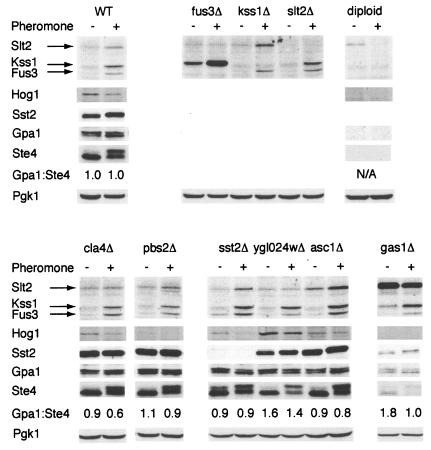
FIG. 7.
Expression of G protein components and activated MAP kinases. For immunoblot detection of phosphorylated MAP kinases, WT cells and the indicated mutant cells were grown to mid-log phase, treated for 1 h with (+) or without (−) 2.5 μM α-factor pheromone as indicated, lysed, and resolved by SDS-PAGE and immunoblotting. Membranes were probed with antibodies to phospho-p44/p42 (to detect phosphorylated Fus3, Kss1, and Slt2), phospho-Tyr (to detect phosphorylated Hog1), Sst2, Gpa1, Ste4, or Pgk1 (loading control), as indicated. Specificity of antibody detection was established using the corresponding gene deletion mutant and/or diploid cells (which do not normally express Sst2, Gpa1, Ste4, or Fus3) (data not shown). Gpa1:Ste4, ratio of Gpa1 and Ste4 expression, as calculated by scanning densitometry; N/A, not analyzed.

