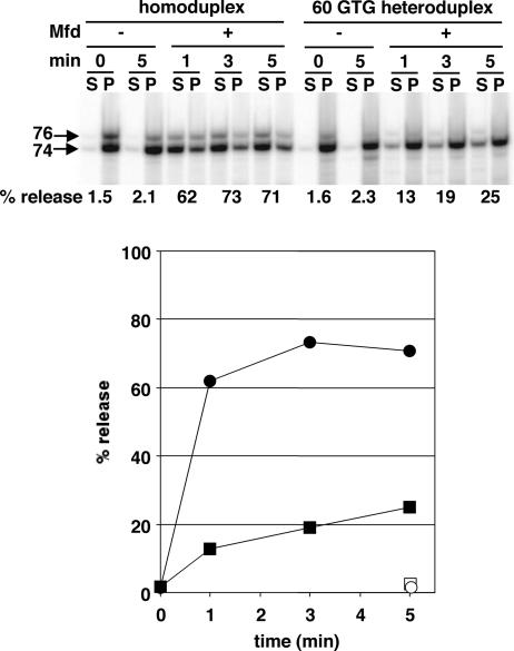
Fig. 3.
Mfd-mediated RNA release from homoduplex and 60 GTG heteroduplex DNA. Each pair of lanes shows a gel analysis of transcripts of stopped elongation complexes affixed through a biotinylated DNA end to magnetic beads; the released or supernatant (S) fraction and retained or pellet (P) fraction after magnetic partitioning is shown. The position of the 60 GTG nontemplate strand substitution is illustrated. The data illustrated in Upper were quantified and are plotted in Lower. Circles, homoduplex DNA; squares, heteroduplex DNA; filled symbols, +Mfd; open symbols, −Mfd.

