
Figure 1.
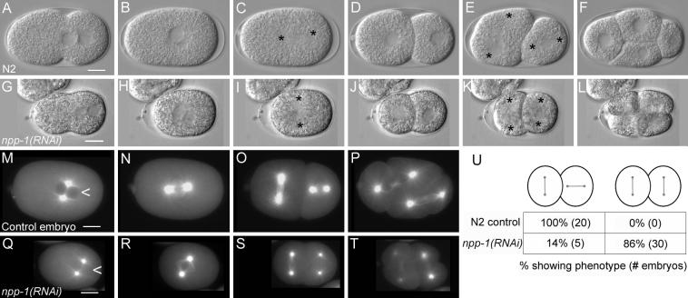
npp-1(RNAi) causes a failed rotation of the mitotic spindle. Panels A-F are a time series of DIC photomicrographs of control embryos (N2). Panels G-L show corresponding time points for npp-1(RNAi) embryos. Anterior is to the left in all embryos. Asterisks mark centrosome positions. No defects in pseudocleavage and pronuclear migration are apparent in npp-1(RNAi) embryos (A versus G). The mitotic spindle fails to rotate properly in P0 and in P1 in npp-1(RNAi) embryos (C, E versus I, K). Panels M-T are photomicrographs of embryos expressing β-Tubulin::GFP at time points comparable to those shown in B,C, E & F. Note the absence of obvious defects in spindle structure. Also note the smaller size of npp-1(RNAi) embryos. Nuclei of npp-1(RNAi) embryos also fail to exclude β-tubulin::GFP (arrowhead in M compared to Q) indicating a failure in nuclear envelope function. A summary table of the P1 spindle orientation defects observed in npp-1(RNAi) embryos is included (U). Bar represents 10 microns.