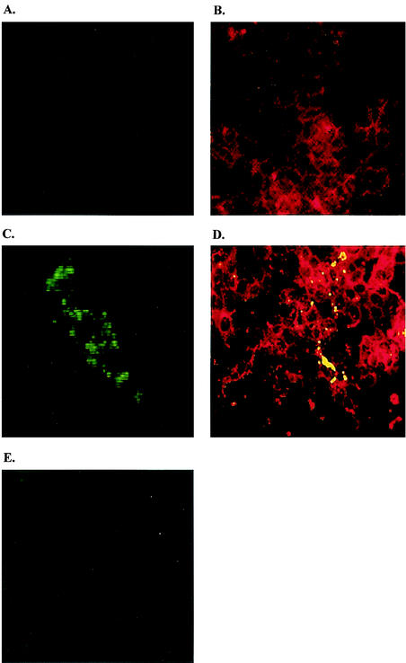
FIG. 3.
Inhibition of HTNV N protein synthesis by ribavirin. (A) Monolayers of Vero E6 cells. (B) Monolayers of Vero cells stained with anti-laminin antibody, followed by TRITC (tetramethyl rhodamine isothiocyanate)-conjugated anti-rabbit immunoglobulin. (C) Vero cells infected with HTNV 76-118 and stained with N mouse monoclonal antibody (EC02-BD01) (22), followed by FITC-conjugated anti-mouse immunoglobulin. (D) Confocal image (overlay) of Vero cells infected with HTNV, stained with anti-laminin TRITC and N mouse monoclonal antibody FITC and observed by using an LSM 510 META microscope (Zeiss). (E) Vero cells infected with HTNV, treated with 24 μg of ribavirin/ml, and stained with N mouse monoclonal antibody. Cells were fixed 72 h after infection and analyzed by immunofluorescence for the presence of HTNV N protein by using a Zeiss axioscope connected to an Orca 100 camera interfaced to a Macintosh computer with Improvision software.

