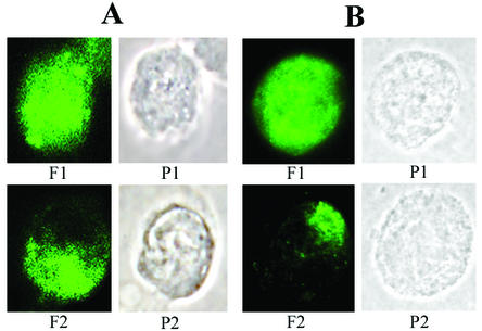
FIG. 1.
Effect of antibodies on glycoprotein distribution in HSV-1-infected HEL (A) and HEp-2 (B) cells. HSV-1-infected cells were incubated with hIgG for 2 h at 37°C, fixed, stained with FITC-labeled secondary antibody, and processed for visualization of HSV-1 surface glycoproteins. The cells were classified according to whether the viral glycoprotein distribution showed uniform staining (1), i.e., the fluorescent label exhibited a uniform cell surface distribution, or was capped (2), i.e., viral glycoproteins formed a large aggregate at one end of the cell. The images are shown in fluorescent (F) and phase-contrast (P) views.

