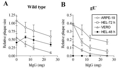
FIG. 4.
Effects of antibodies on cell-to-cell spread of wild-type (A) and gE− (B) HSV-1 in different cell types. The indicated cells were infected with virus and subsequently cultured for 48 h in the presence of the indicated concentration of hIgG. In HEL cells infected with gE− HSV-1, no plaques were discernible at 48 h postinfection in the absence of antibody but were visualized and quantifiable at 72 h postinfection. Plaque measurements were made at 48 h postinfection unless indicated as 72 h. Twenty to thirty plaques were used to estimate average plaque size under each condition. Results shown are representative of several independent sets of analyses.

