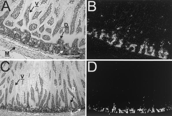
FIG. 1.
In situ hybridization analysis of enJSRV mRNA expression in Peyer's patch tissue collected from the small intestine of fetal lambs (gestation day 120). Cross sections of different regions of the small intestine from sheep fetuses were hybridized with α-35S-labeled antisense ovine enJSRV cRNA probes. Protected transcripts were visualized by liquid emulsion autoradiography for 1 week and imaged under bright-field or dark-field illumination. (A) Bright field of jejunal Peyer's patch tissue stained with hematoxylin. Numerous lymphoid aggregates (L) are visible between the muscolaris externa (M) and the overlying mucosal epithelium (V). Domes (D) are also visible. (B) In situ hybridization reveals a high degree of enJSRV expression that localizes to cells within the lymphoid aggregates of the jejunal Peyer's patches. (C) Bright field of ileal Peyer's patch tissue stained with hematoxylin. Small lymphoid aggregates are visible between the muscularis externa (M) and the overlying mucosal epithelium (V). (D) In situ hybridization reveals enJSRV RNA expression that is localized to cells within the lymphoid aggregates of the Peyer's patch. Bar, 150 μm.

