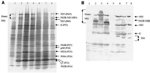
FIG. 3.
Analysis of TBEV-specific proteins by PAGE (A) and Western blotting (B). Infected PS cell monolayers were lysed 48 h postinfection for Sof and Za viruses and 72 h postinfection for Vs virus. Proteins were separated by electrophoresis on 7 to 15% polyacrylamide gels. For Western blotting, the proteins were transferred onto a nitrocellulose membrane and revealed by interaction with anti-TBEV antibodies. Lanes 1 and 5 contained mock-infected PS cells; the other lanes contained PS cells infected with Sof (lanes 2 and 6), Za (lanes 3 and 7), and Vs (lanes 4 and 9) viruses. Samples 1 to 4 were unheated and samples 5 to 8 were heated for 1 min at 95°C. The virus-encoded proteins and their masses are specified.

